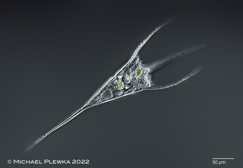
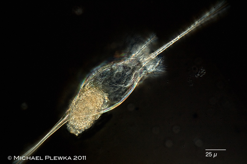
| Kellicottia longispina; specimen from (7); dorsoventral view. |
| |
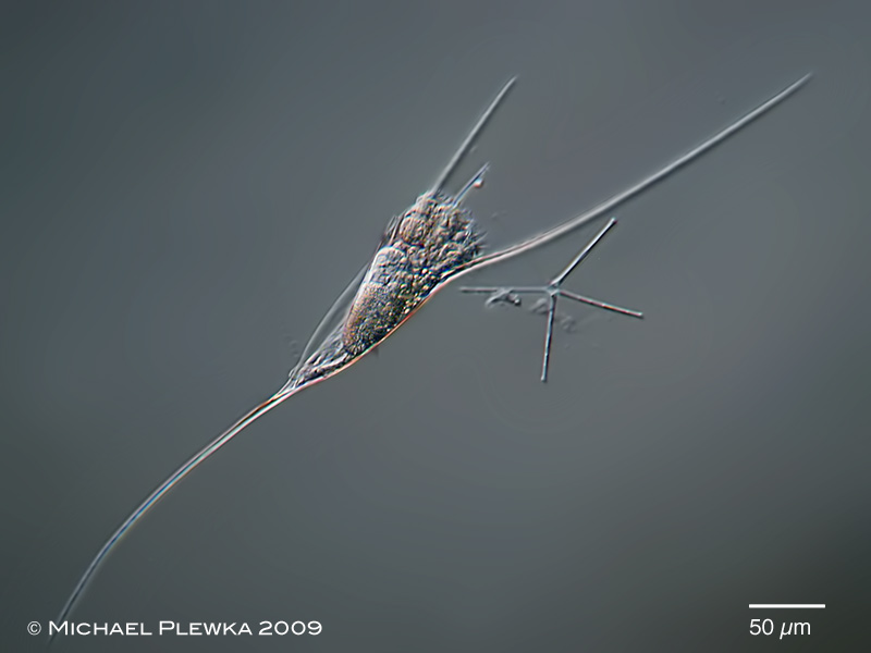 |
| Kellicottia longispina: the long spines of this plankton rotifer act as help for floating in the water. The same principle applies to the diatom Asterionella which is also visible in this image. Likewise the spines act as protection against predators like the big rotifer Asplanchna. Kellicottia is simply too bulky to be ingested by Asplanchna. (1) |
| |
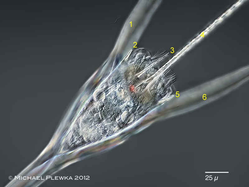 |
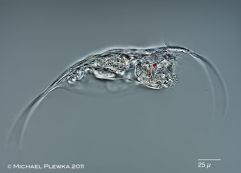
| Kellicottia longispina: dorsoventral view; this species has 6 anterior spines, which is in contrast to Kellicottia bostoniensis (4 anterior spines) |
| |
 |
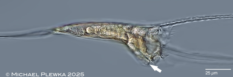
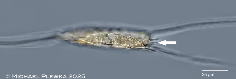
| Kellicottia longispina: ventral view, corona retracted; focal plane on the two projections at the anterior margin of the ventral plate (arrows), which seem to "guide" the two ventral lobes of the corona. (7) |
| |
 |
 |
| Kellicottia longispina: lateral view; the above mentioned projections are flexible: in the upper image the arrow points to one of these projections. Both are bent caudally when the corona is expanded. In the lower image the same projection is bent dorsally (arrow) when the corona is retracted. Both images stills from a video showing this movement. (8) |
| |
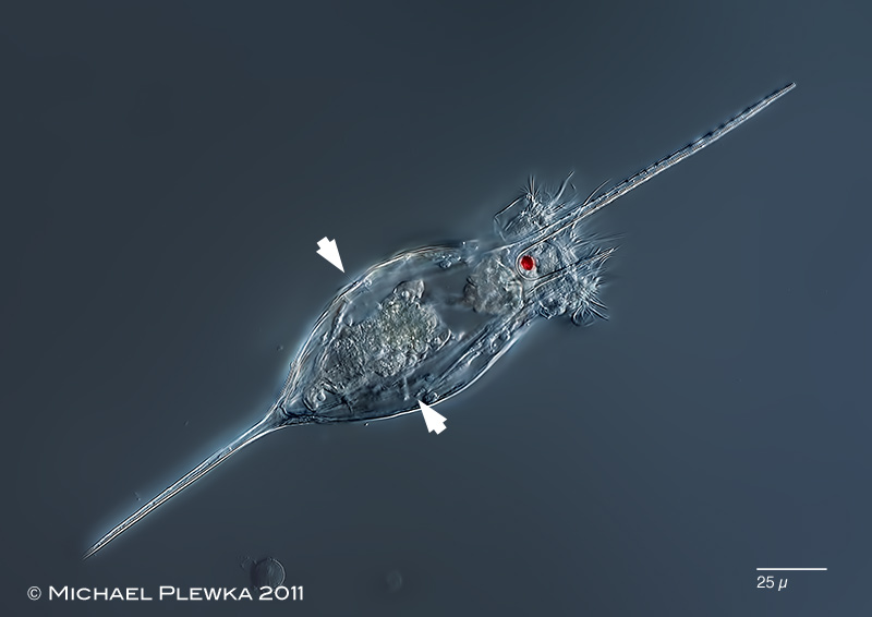 |
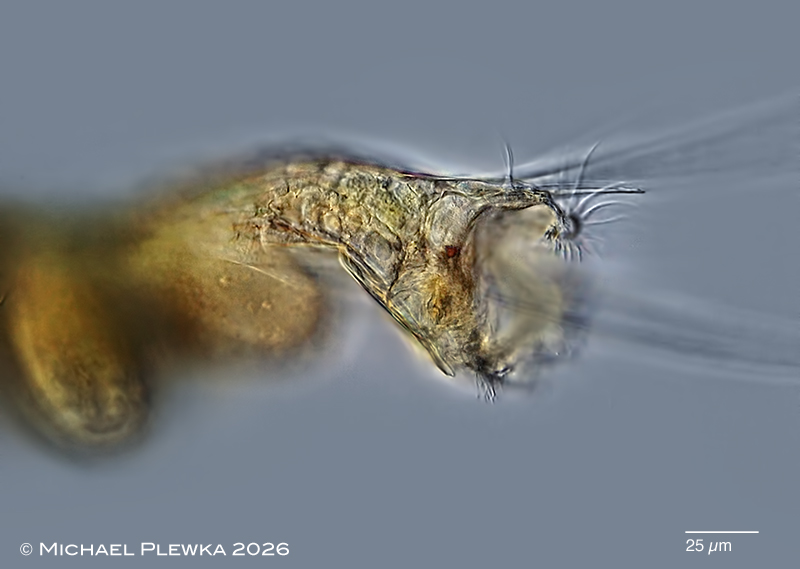
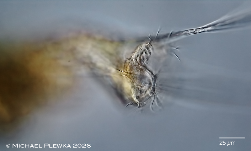
| Kellicottia longispina, dorsal view, focus plane on the red eyespot. The arrowheads point to the lateral antennas. (3); |
| |
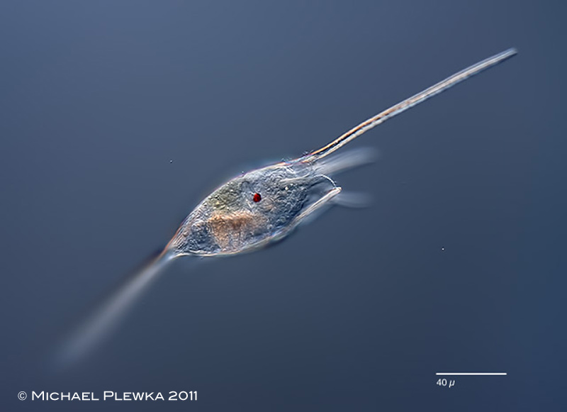 |
| Kellicottia longispina; the length of the spines vary with environmental conditions. (see first image). (3) |
| |
|
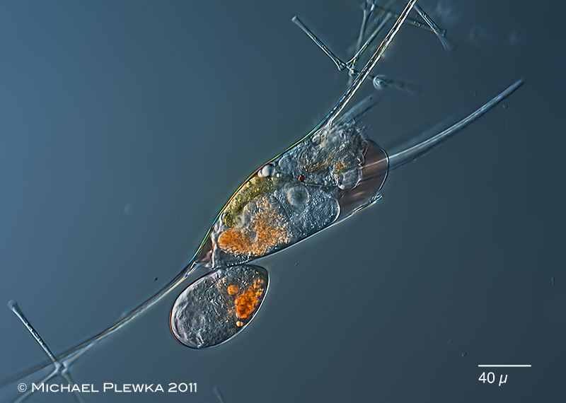
| Kellicottia longispina, specimen with amictic egg. (7) |
| |
 |
| Kellicottia longispina, specimen with resting egg. The shell is sculptured like the one from Keratella. (3) |
| |
 |
| Kellicottia longispina; recently hatched specimen with (still) curved spines. (4) |
| |
 |
 |
| Kellicottia longispina; two images of specimen in lateral view. Upper image: optical median transect; focal plane on the eyespot, dorsal antenna and the bristles at the median dorsal lobe. Lower image: focal plane on the right lateral lobe. (8) |
| |
 |
 |
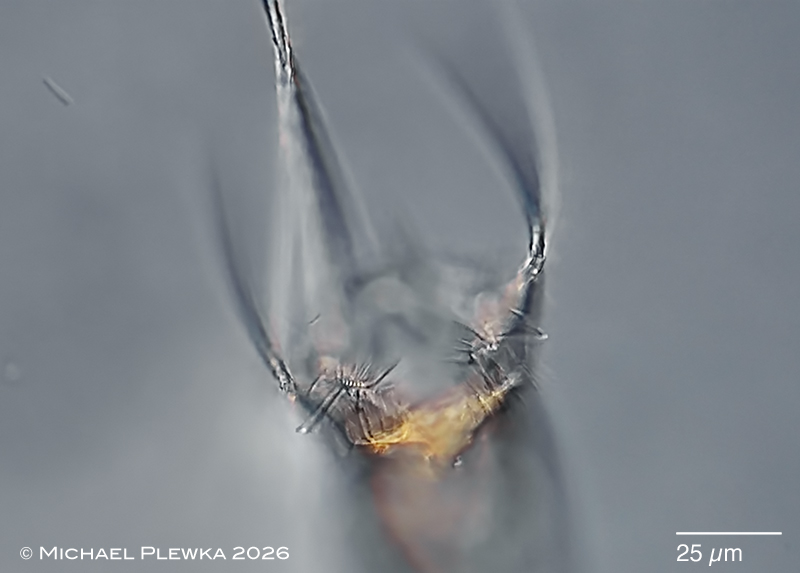 |
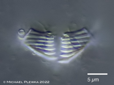
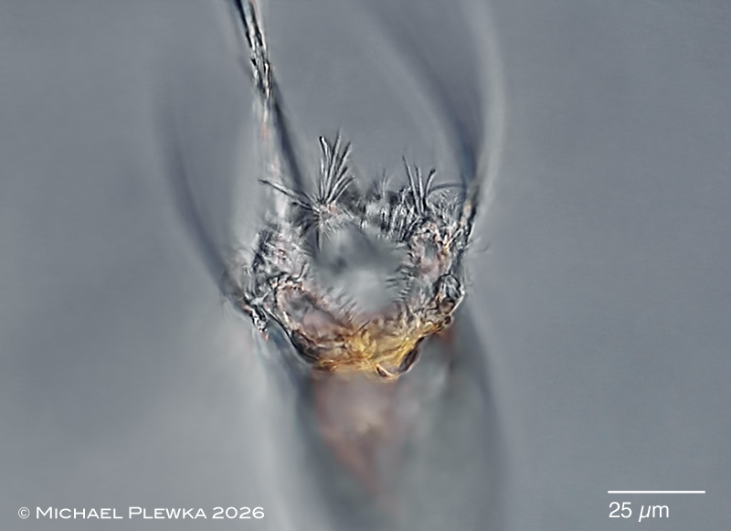
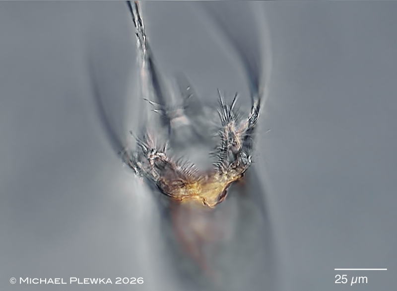
Kellicottia longispina; three images of the corona in ventral view (about 45 degrees of the longitudinal axis).
Upper image: focal plane on the dorsal median lobe, the rim of which is fitted with cilia that have clockwise metachronal waves.
Mid image: focal plane on the median lobes which are fitted with bristles. Also visible are the cilia of the buccal field.
Lower image: focal plane on the ventral lobes, each with a bunch of bristles. Also visible is the "lower lip" with cilia with laeoplectic metachronal waves in clockwise direction.(9) |
| |
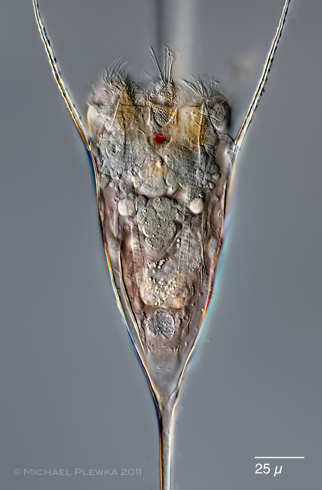 |
| Kellicottia longispina; another specimen (28.05.2011), dorsoventral view. Focus plane on the red eyespot and the 3-lobed corona (Euchlanis-type). The body fluid is reddish similar to one from Gastropus stylifer. 2 of the 4 transversely striated (retractor) muscles are visible. (28.05.2011).(6) |
| |
|
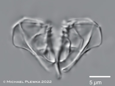
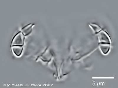
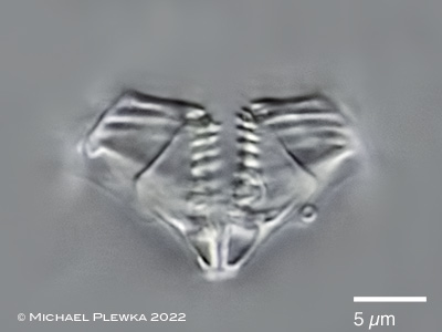
| Kellicottia longispina, malleate trophi of macerated specimen. Upper left:focus plane on the manubria; upper right: manubria in optical transect. Lower left: focus plane on the unci; lower right: focus plane on the rami. (7) |
|
| |
| |
|
|
| |
| |
|
|
| Location: Glörtalsperre / NRW (1);(6); Heilenbecker Talsperre (3); NSG Heiliges Meer, Großes HM (4); |
| Habitat: plankton (1); (2); (3); (4);(6) |
| Date: 06.04.2009(1); 7.4.2009 (2); 16.01.2011 (3); 21.03.2011 (7); 16.04.2011 (4); 28.05.2011 (6) |
|
|